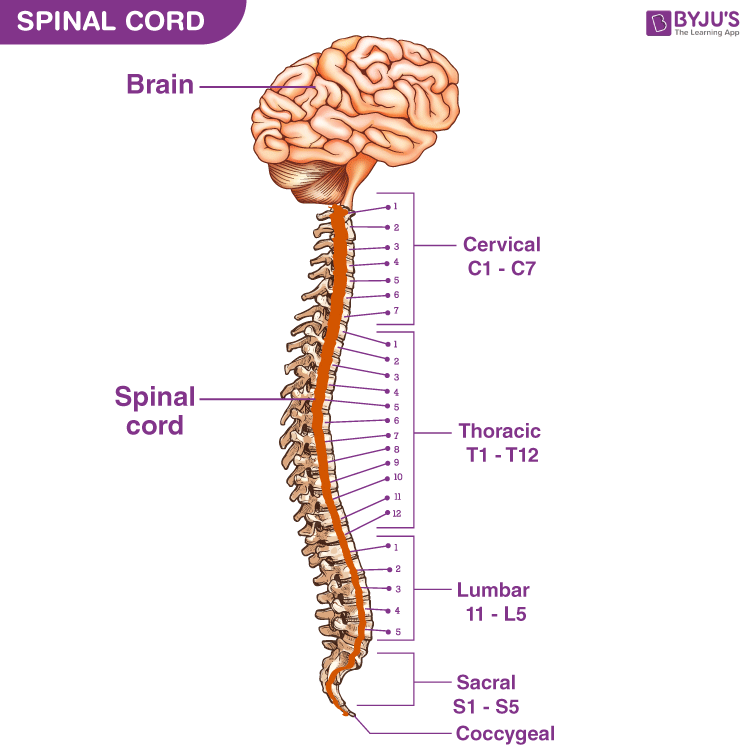

Removing irritation and restoring balance to the nervous system enhances the body’s capacity to heal. Spine Anatomy, Diagram & Pictures Body Maps Human body Skeletal System Spine Spine The spinal cord begins at the base of the brain and extends into the pelvis. This is why chiropractic can have a beneficial effect on certain conditions such as asthma, indigestion, constipation and premenstrual syndrome to name a few. The spine is a very complex mechanical structure that is highly flexible yet very strong and stable. When the nerves that supply your organs, like the stomach, heart and lungs are irritated, these organs cease to function at their optimal level. Browse 899 spine diagram stock photos and images available, or search for back pain or chiropractor to find more great stock photos and pictures. Exercises can strengthen the core muscles that support the spine and. The disks that cushion vertebrae may compress with age or injury, leading to a herniated disk. The vertebral column (also known as the backbone or the spine), is a column of approximately 33 small bones, called vertebrae. The spine supports your body and helps you walk, twist and move.

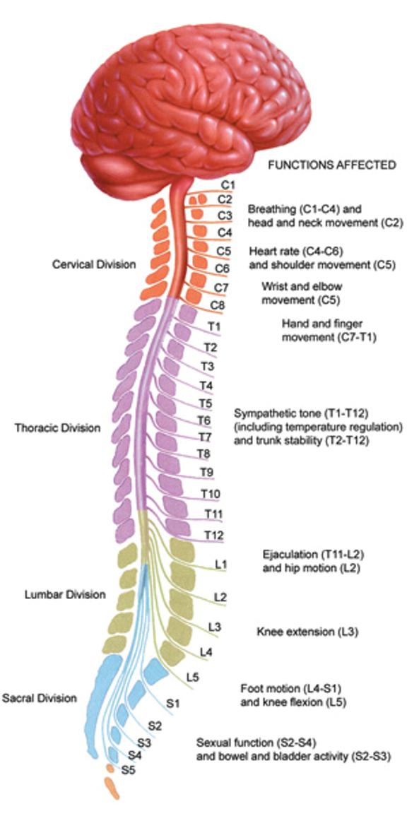
It is also great for restoring proper nerve function to the rest of your body that you don’t think about. The lumbo-sacral spine includes: Lumbar vertebrae: Numbered L1 through L5, these odd-shaped vertebrae signal the end of the typical bones of the spinal column. A typical vertebra consists of an vertebral body at the front (anterior) and a vertebral (neural) arch at the back (posterior). Key parts of your spine include vertebrae (bones), disks, nerves and the spinal cord. Simply line up the “Vertebral Level” with the “Possible Symptoms” and you will see some surprising connections of symptoms that relate to your spine.Ĭhiropractic is great for back, neck and extremity pain. T1-T12 is the UPPER BACK /rib cage area, and.Damage to the nerves, nerve roots or spinal cord may result in symptoms such as pain, tingling, numbness and weakness, both in and around the damaged area and in the extremities.On the chart below you will see 4 Columns (Vertebral Level, Nerve Root, Innervation, and Possible Symptoms). The nerves also carry electrical signals back to the brain, creating sensations. Those in the cervical spine, for example, extend to the upper chest and arms those in the lumbar spine the hips, buttocks and legs. The nerves in each area of the spinal cord are connected to specific parts of the body. Inside this sac is spinal fluid, which surrounds the spinal cord.

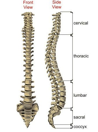
The spinal cord is covered by a protective membrane called the dura mater, which forms a watertight sac around the spinal cord and nerves. From the side, our three spinal column segments form. Because of its appearance, this group of nerves is called the cauda equina – the Latin name for “horse’s tail.” The nerve groups travel through the spinal canal for a short distance before they exit the neural foramen. The cervical, thoracic and lumbar spine make up the three main segments of the spinal column, or backbone. Its anatomy is extremely well designed, and serves many functions, including: Movement Balance Upright posture Spinal cord protection Shock absorption.

The spinal cord ends by diverging into individual nerves that travel out to the lower body and the legs. It consists of 33 vertebrae, or backbones, stacked atop one another, from the tailbone to the. The spinal cord extends from the base of the brain to the area between the bottom of the first lumbar vertebra and the top of the second lumbar vertebra. The spine is both relatively simple and amazingly complex.


 0 kommentar(er)
